 LymyVoyage
Dental AI X-Ray Reports: How Often Is a Flagged Problem Real?
Research & verification
We independently research information from publicly available sources. Claims are cross-checked against multiple sources where possible. When sources disagree, the disagreement is reported rather than resolved by assumption. LymyVoyage does not provide medical advice or make individual treatment recommendations.
LymyVoyage finding
Disputed -- sources materially disagree.
Two independent studies put Diagnocat's specificity for decay on panoramic X-rays at 98% and 47%, using different images, reference readers and software years; neither explains the gap.
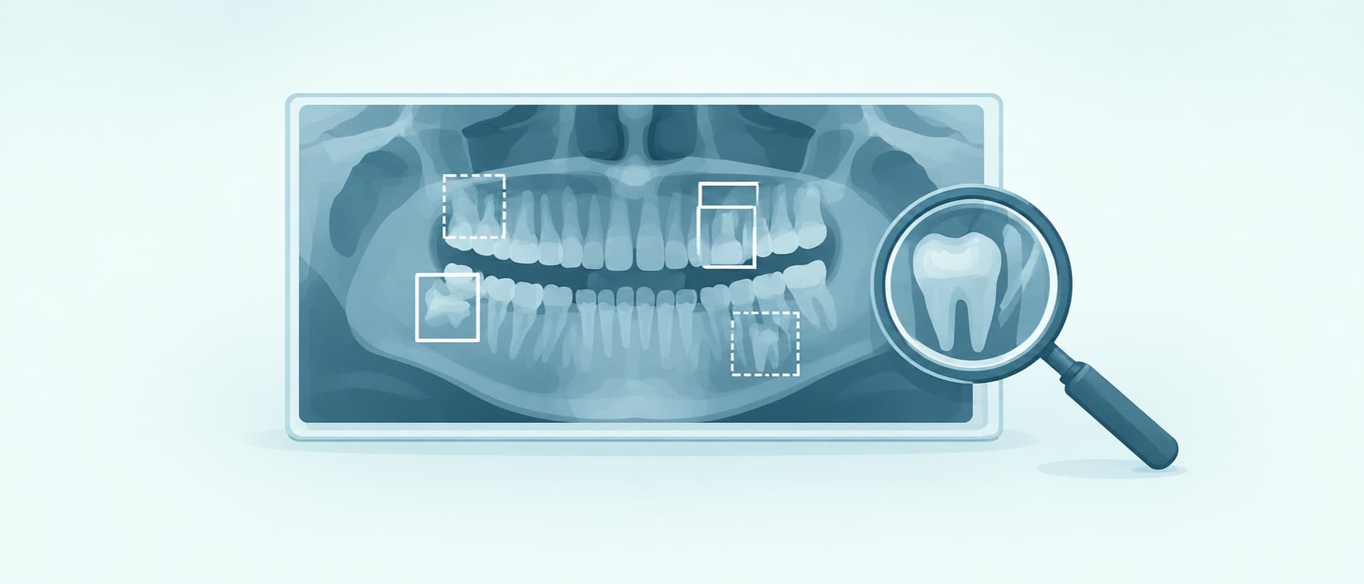
A dentist can now hand you a printed report in which software has marked every tooth on your X-ray. Diagnocat, one of the companies selling these tools, tells patients its report is "a reliable second opinion powered by advanced AI technology."1 Its own blog describes clinics running international patients' scans through the software before they travel, then sending back a report with "potential treatment needs" and suggested treatment order.2
So the report can end up shaping a treatment plan before anyone has looked in your mouth. That makes one question worth answering with published numbers rather than marketing: when dental AI flags a problem on your X-ray, how often is the problem real?
Dental AI now marks problems on your X-ray and prints a report you can take home. One maker calls it "a reliable second opinion." How often is a flagged problem real?
| Tool | Cleared in the US to find | X-ray type | Sensitivity | Specificity or false marks | Decision |
|---|---|---|---|---|---|
| Overjet Caries Assist | Decay | Bitewing | 72.0% | 98.1% per tooth surface | May 20223 |
| Pearl Second Opinion CS | Decay | Periapical | 88% per lesion | +0.16 false marks per image when dentists used it | June 20254 |
| Pearl Second Opinion Panoramic | Decay, dark areas at the root tip, impacted wisdom teeth | Panoramic | 77% for decay | Not given in the summary reviewed | Nov 20255 |
| Diagnocat | Dark areas at the root tip only | Cone-beam CT (3D scan) | 85.4% | 99.1% | Jan 20266 |
What a US clearance actually covers
Each US Food and Drug Administration (FDA) clearance reviewed here names specific findings on one image type. Each is for use alongside a dentist, who reads the X-ray with the software's marks on screen. None is cleared to diagnose on its own.
Overjet's caries tool is the clearest example. In the company's FDA test, it found 72.0% of decayed surfaces on 352 bitewing X-rays.3 It wrongly flagged 1.9% of healthy surfaces, a specificity of 98.1%. When 13 US dentists read the same images with and without it, their sensitivity rose from 57.9% to 76.2%. Their specificity slipped from 99.3% to 98.4%.
A 98% figure sounds close to perfect, but it is counted per tooth surface, and Overjet's test set held about 20 surfaces per X-ray. At that rate, a set of healthy teeth would collect roughly one false flag for every two or three bitewing images. That figure is this publication's arithmetic from the FDA summary, not a number Overjet publishes.
Pearl reports its tools differently. Its periapical caries tool found 88% of lesions in a test of 1,250 images.4 When 21 dentists used it, the summary reports a "slight increase" of 0.16 false marks per image. Pearl's panoramic tool, cleared in November 2025, found 77% of decay on panoramic films.5
Diagnocat's US clearance is narrower than its report
The FDA's database lists two clearances under Diagnocat's US company, DGNCT LLC.7 One, from September 2025, is a viewer for 3D scans. Its summary says it makes no diagnostic claims.8 The other, from January 2026, is for one finding: dark areas at the tips of roots on cone-beam CT, a 3D dental scan.6 On that task it scored 85.4% sensitivity and 99.1% specificity.
That is a much smaller scope than the report patients see. Diagnocat's patient page promises "a clear description of each tooth, warning signs" and dentist comments.1 The same page adds that features "may not be available or approved for use in all regions."
By comparison, the same FDA database lists 13 clearances under Overjet's name.9 It lists six detection clearances for Pearl, including the panoramic tool.10 So a patient abroad may receive a full-mouth Diagnocat report, of which the US has reviewed one finding on one scan type. The rules in the clinic's own country are a separate question this article did not check.
Independent tests of Diagnocat disagree
Panoramic X-rays are the wide films that show the whole mouth in one image. Diagnocat's dental-tourism blog lists them among the scans it analyses.2 Three published studies tested Diagnocat on them, and their results point in different directions.
| Study | Images | Who set the right answer | Decay: sensitivity | Decay: specificity |
|---|---|---|---|---|
| Zadrożny et al., Warsaw, 202211 | 30 | The authors' reference reading | 44.5% | 98.2% |
| Orhan et al., 202312 | 100 | Three radiologists | Low (agreement score 0.365) | Not reported in the abstract |
| Mema et al., 202513 | 104 | Three dentists | Within 77-96% | 47% |
The 2022 Warsaw study is small, and its authors declared no conflict of interest.11 It found Diagnocat cautious about decay: the software missed more than half of it, but almost never flagged a healthy tooth. It also missed 61% of root-tip lesions.
The 2025 study, published in Applied Sciences, found nearly the opposite pattern.13 Diagnocat caught 99.2% of conditions the experts marked. But its overall specificity was 8.7%, which the authors read as "a tendency to overdiagnose." For decay, specificity was 47%. Its agreement with the experts on decay scored −0.15 on a scale where zero means chance-level agreement.
Both figures are published, peer-reviewed and specific. They cannot both describe the same tool in the same conditions. The studies used different image sets, different reference panels and different years of the software, and none of them explains the gap. This article does not pick between them.
The 2023 study is the most favourable. It reported "almost perfect agreement" on most findings, but low sensitivity for decay and root-tip lesions.12 Eight of its 12 authors list Diagnocat Inc. as their employer. Its lead author is a scientific adviser to the company, according to the paper's own disclosure.
Precision: the number that answers your question
Accuracy studies usually report two numbers. Sensitivity is the share of real problems the software finds, and specificity is the share of healthy teeth it correctly leaves alone. Neither answers the question a patient holding a report has. That question is precision: of the spots the software flagged, how many were real?
Precision depends on how common the problem is. When most teeth are healthy, even a small false-flag rate can put many wrong marks next to a few real ones.
A 2024 study of 600 panoramic X-rays shows the effect, using a different product from the ones above.14 It tested Carestream Dental's CS Imaging software against two human readers. For root-tip lesions, the software scored 99.2% specificity. Yet only 72.5% of its root-tip flags matched the readers, so roughly one in four did not.
Most of the Diagnocat studies above report sensitivity and specificity, not precision. A patient usually cannot look up the figure that matters most for the tool their clinic uses.
Better detection can also mean more drilling
Even a tool that finds decay accurately can change what happens next. A randomised trial at Charité hospital in Berlin tested this directly.15 Twenty-two dentists read bitewing X-rays with and without an AI tool called dentalXrai Pro.
With AI help, their sensitivity rose from 0.72 to 0.81, while their specificity did not change significantly. The gain came from early decay in the outer enamel, not from deeper lesions.
In a randomised trial of 22 dentists, AI support made them find more early decay and also choose invasive treatment more often.15
Both non-invasive and invasive treatment decisions rose. The authors concluded that AI "may also increase invasive treatment decisions." The software maker was founded by two of the trial's authors, Falk Schwendicke and Joachim Krois.16 Their trial still published the downside.
The case for the tools
The evidence does not say dental AI is unreliable. A 2025 umbrella review in PLoS One pooled 20 studies of AI decay detection.17 It found pooled sensitivity of 85% and specificity of 90%.
In the US clearance studies above, dentists found more disease with the software than without it.
The same review also sets limits on its own numbers. Only 20 of 137 studies it found reported enough data to pool. The decay rate in the test sets was 27%, which the authors call lower than in real communities. They found signs of publication bias.
At that 27% rate, the review calculates that a positive AI result means a 79% chance of real decay.17 Put the other way round, about one positive flag in five would be wrong.
What the numbers leave out
Who decided the right answer. Every accuracy figure above measures the software against human readers. The Warsaw and 2025 studies used different panels and got opposite results. None of the studies here checked a flag against what was found when the tooth was opened.
The image you actually had. A clearance for bitewings says nothing about a panoramic film. Diagnocat's detection clearance in the US covers 3D scans, not panoramic films.
Who reads the report. The US clearances assume a dentist reviews each mark. A report emailed to a patient, or used to build a quote abroad, may skip that step. None of the sources here measures how often that happens.
Your clinic's version and settings. Diagnocat assigns each finding a likelihood score from 0 to 100%.13 Where a clinic sets the cut-off changes how many flags appear. Patients are not told the setting.
How to use an AI report on your X-ray
Ask which software produced the report, and which image type it read. Then check whether that tool is cleared for that finding on that image. In the US, the FDA's 510(k) database is public and searchable by company name.
Ask the dentist to point to each flagged spot and say whether they agree with it, because a flag they do not stand behind should not appear on your quote.
For any flag of early decay, ask whether it can be watched or treated without drilling. That is the decision the Berlin trial showed AI can push the wrong way.
Before crowns, veneers or root canals, get the same X-ray read by a dentist who is not selling the treatment. An AI report can be one of the inputs to that second look. It cannot be the second look.
Footnotes
-
https://diagnocat.com/en/solutions/patients — Diagnocat, "Diagnocat for patients" (no publication date stated). Accessed Oct 12, 2026. ↩ ↩2
-
https://diagnocat.com/blog/ai-revolution-in-dental-tourism-transforming-smiles-across-borders — Diagnocat, "AI Revolution in Dental Tourism: Transforming Smiles Across Borders" (published Aug 6, 2024). Accessed Oct 12, 2026. ↩ ↩2
-
https://fda.innolitics.com/device/K212519 — US Food and Drug Administration, 510(k) summary K212519, "Overjet Caries Assist" (decision May 10, 2022), via Innolitics' FDA document mirror. Accessed Oct 12, 2026. ↩ ↩2
-
https://fda.innolitics.com/device/K243234 — US Food and Drug Administration, 510(k) summary K243234, "Second Opinion CS," Pearl Inc. (decision Jun 12, 2025), via Innolitics' FDA document mirror. Accessed Oct 12, 2026. ↩ ↩2
-
https://fda.innolitics.com/device/K250525 — US Food and Drug Administration, 510(k) summary K250525, "Second Opinion Panoramic," Pearl Inc. (decision Nov 14, 2025), via Innolitics' FDA document mirror. Accessed Oct 12, 2026. ↩ ↩2
-
https://fda.innolitics.com/device/K252934 — US Food and Drug Administration, 510(k) summary K252934, "Diagnocat," DGNCT LLC (decision Jan 15, 2026), via Innolitics' FDA document mirror. Accessed Oct 12, 2026. ↩ ↩2
-
https://api.fda.gov/device/510k.json?search=applicant:%22DGNCT%22 — US Food and Drug Administration, openFDA 510(k) database, query for applicant "DGNCT" (two records returned). Accessed Oct 12, 2026. ↩
-
https://fda.innolitics.com/device/K251072 — US Food and Drug Administration, 510(k) summary K251072, "Segmentron Viewer," DGNCT LLC (decision Sep 9, 2025), via Innolitics' FDA document mirror. Accessed Oct 12, 2026. ↩
-
https://api.fda.gov/device/510k.json?search=applicant:%22Overjet%22&limit=40 — US Food and Drug Administration, openFDA 510(k) database, query for applicant "Overjet" (13 records returned). Accessed Oct 12, 2026. ↩
-
https://api.fda.gov/device/510k.json?search=applicant:%22Pearl%22+AND+product_code:MYN&limit=30 — US Food and Drug Administration, openFDA 510(k) database, query for applicant "Pearl" under detection product code MYN (six records returned). Accessed Oct 12, 2026. ↩
-
https://pmc.ncbi.nlm.nih.gov/articles/PMC8774336/ — Zadrożny Ł. et al., "Artificial Intelligence Application in Assessment of Panoramic Radiographs," Diagnostics (published 2022). Accessed Oct 12, 2026. ↩ ↩2
-
https://pubmed.ncbi.nlm.nih.gov/37799743/ — Orhan K. et al., "Determining the reliability of diagnosis and treatment using artificial intelligence software with panoramic radiographs," Imaging Science in Dentistry (published Aug 2023). Accessed Oct 12, 2026. ↩ ↩2
-
https://doi.org/10.3390/app15179790 — Mema H. et al., "Application of AI-Driven Software Diagnocat in Managing Diagnostic Imaging in Dentistry: A Retrospective Study," Applied Sciences (published Sep 6, 2025); abstract read via the Crossref record. Accessed Oct 12, 2026. ↩ ↩2 ↩3
-
https://pmc.ncbi.nlm.nih.gov/articles/PMC11595016/ — Turosz N. et al., "Evaluation of Dental Panoramic Radiographs by Artificial Intelligence Compared to Human Reference: A Diagnostic Accuracy Study," Journal of Clinical Medicine (published 2024). Accessed Oct 12, 2026. ↩
-
https://pubmed.ncbi.nlm.nih.gov/34656656/ — Mertens S. et al., "Artificial intelligence for caries detection: Randomized trial," Journal of Dentistry (published Dec 2021). Accessed Oct 12, 2026. ↩ ↩2
-
https://dha.bihealth.org/ai-in-the-fight-against-cavities-bih-digital-health-accelerator-supports-spin-off-of-the-start-up-dentalxrai/ — Berlin Institute of Health Digital Health Accelerator, "AI in the fight against cavities" (published Aug 10, 2020). Accessed Oct 12, 2026. ↩
-
https://pmc.ncbi.nlm.nih.gov/articles/PMC12349118/ — Arzani S. et al., "Examining the diagnostic accuracy of artificial intelligence for detecting dental caries across a range of imaging modalities: An umbrella review with meta-analysis," PLoS One (published 2025). Accessed Oct 12, 2026. ↩ ↩2
Evidence at a glanceMethodology
| Statement | Source | Tier | Date | Status |
|---|---|---|---|---|
| In its FDA test of 352 bitewing X-rays, Overjet Caries Assist found 72.0% of decayed surfaces and wrongly flagged 1.9% of healthy surfaces (98.1% specificity) | FDA 510(k) summary (via Innolitics mirror)3 (Archived copy) | 1 | 2022-05-10 | Approximate |
| Diagnocat's only US detection clearance (K252934, January 2026) covers periapical radiolucency on cone-beam CT; its other US clearance (K251072) is a viewer that makes no diagnostic claims | FDA 510(k) summary (via Innolitics mirror)6 (Archived copy) | 1 | 2026-01-15 | Supported |
| Pearl's Second Opinion CS found 88% of caries lesions on periapical X-rays, and dentists using it marked 0.16 more false positives per image | FDA 510(k) summary (via Innolitics mirror)4 (Archived copy) | 1 | 2025-06-12 | Approximate |
| Diagnocat's specificity for caries on panoramic X-rays was 98.2% in a 30-image Warsaw study (2022) and 47% in a 104-image study (2025) | Peer-reviewed diagnostic accuracy studies11 | 1 | 2022; 2025-09-06 | Disputed |
| In the 2025 Applied Sciences study, Diagnocat's overall sensitivity was 99.2% and overall specificity 8.7%, which the authors describe as a tendency to overdiagnose | Peer-reviewed diagnostic accuracy study13 | 1 | 2025-09-06 | Approximate |
| Eight of the 12 authors of the 2023 Orhan et al. Diagnocat study list Diagnocat Inc. as their affiliation, and the lead author is a scientific adviser to the company | PubMed record with author affiliations and conflict-of-interest statement12 | 1 | 2023-08-02 | Approximate |
| In a randomised trial of 22 dentists, AI support raised caries sensitivity from 0.72 to 0.81 without a significant change in specificity, and increased invasive treatment decisions | Peer-reviewed randomised trial15 | 1 | 2021-12 | Approximate |
| An umbrella review of 20 studies pooled AI caries-detection sensitivity at 0.85 and specificity at 0.90, with a 79% post-test probability of caries after a positive result | Peer-reviewed umbrella review with meta-analysis17 | 1 | 2025 | Approximate |
| At Overjet's reported 1.9% per-surface false-positive rate and about 20 surfaces per bitewing, healthy teeth would collect roughly one false flag per two to three images | Calculated by this publication False-positive rate (1 - 0.981) multiplied by surfaces per image (7,129 surfaces / 352 images = 20.3), giving about 0.38 false flags per image if all surfaces were healthy. | 1 | -- | Approximate |
Enjoyed this? Get more like it
Occasional emails, no spam -- just new articles as they're published.
More from AI & Medical Travel



Comments
Loading comments…